
Research has shown that magnetic resonance imaging (MRI) for back pain remains controversial because a considerable number of patients may be classified incorrectly by MRI for lumbar disc herniation and spinal stenosis. These studies suggests that MRI may not be pointing to the correct cause of traditionally attributed back pain–herniated disc and spinal stenosis. Further, there is more confusion being created because the medical research does not agree. Here is why a physical examination should be considered a main diagnostic tool.
As with other joint problems, I will often get emails that are simply an MRI report. The MRI tells me that the person has a disc herniation at L1 or L2 or L4 or L5. Then the report will describe varying degrees of degenerative disc disease. The email will end with “can you help?” That answer would be easier if there was some more information included in that email.
At no point in the email did the person say how bad their pain was, what type of limitations they had, or how their back problem was affecting their ability to work or be active. Information as simple as knowing how someone’s back feels today is good information to have when trying to determine if our treatments can help them instead of surgery.
For some people the MRI of their back pain has terrified them.
It is not the emailers fault for excluding this information. For some people, they have been trained that the MRI has captured the image of what is causing their pain and this image can be used as a roadmap or baseline to help doctors plot out a surgical path now or in the near future.
I get the MRI report because these people are exploring ways to avoid that surgery. For some people the MRI has terrified them.
Too many MRIs? Not enough MRIs? patients and doctors are confused by what is an appropriate MRIs
There was a paper published in the Journal of physiotherapy (18) It suggested that people with low back pain are centered around their desire for a diagnosis, potentially contributing to expectations for and overuse of imaging. People with low back pain expressed a strong desire for clear, consistent and personalized information on prognosis, treatment options and self-management strategies, related to healthcare and occupational issues.”
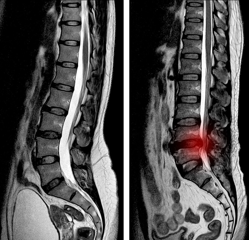
There are many structures in the lower back that can cause severe pain. Not all are clear on an MRI
There are many structures in the lower back that can cause severe pain. These include muscles, ligaments, tendons, bones, joints and discs. For example, the outer rim of the disc can be a significant source of back pain due to its rich nerve supply and tendency toward injury.
During our body’s development, there is a great deal of overlap of nerve supply to all these structures. This makes it nearly impossible for the brain to distinguish between injury to one structure versus another.
For example, a torn or herniated disc can feel identical or similar to a bruised muscle or ligament injury. This is where an examination into the cause of the pain is important.
Axial or mechanical back pain
This pain can be a very sharp to a dull ache. It may occur all the time, or it may come and go. It also varies in intensity from very mild to extremely severe. One patient may report that his/ her lower back is only sore when having been seated for a long time, or after working in the garden. Another patient may report severe, debilitating pain and need assistance to walk or stand, or even to get up from a sitting position. While one patient is perfectly straight, the other is bent over and locked in a crooked posture. The one thing that is common in these conditions is that the pain is restricted to the lower back area.
Referred pain
Here, patients complain of having an achy, dull type of pain that seems to move around. The discomfort comes and goes and varies in intensity. This achy pain starts in the low back area and commonly spreads into the groin, Buttock and upper thighs.
Radicular pain
In this case, the pain is described as deep and usually constant. It follows the nerve down the leg and is often accompanied by numbness or tingling and muscle weakness.
The most common example of this type of problem is the sciatic pain that radiates along that sciatic nerve – down the back of the thigh and calf into the foot. This type of pain is caused by injury to a spinal nerve. Some of the possible causes of this are a disc protrusion or bulge, arthritic changes or a narrowing of the opening through which the nerve exits.
So with all this pain a person can easily think the worst. This is shown in the medical literature.
“The catastrophization effects of an MRI report on the patient”
In July 2021 a study (1) was published that suggested that possibility of a catastrophizing effect on the patient. What does that mean? Here is what the paper’s authors said:
“Inappropriate use of MRI leads to increasing interventions and surgeries for low back pain. (The researchers) probed the potential effects of a routine MRI report (how the report was explained to the patient) on the patient’s perception of his spine and functional outcome of treatment.”
This is how the study was conducted:
- In Phase I – 44 low back pain patients were randomized to Group A who had a factual explanation of their MRI report or Group B, who were reassured that the MRI findings showed normal changes.
- In Phase-II, clinical reporting was developed, (an alternative way of talking to patients) avoiding potential catastrophizing terminologies.
- In Phase-III, 20 MRIs were reported by both routine and clinical methods. The effects of the two methods were tested on four categories of health care professionals who read them blinded on their assessment of severity of disease, possible treatment required, and the probability of surgery.
In summary: Two groups of low back pain patients. One group (Group A) had their MRI read to them as you would get any MRI report. The second group (Group B) had their MRI report explained to them minimizing terminology that would concern the patient and all the while reassuring the patient that the MRI interpretation were normal of aging and degenerative changes.
- Results: “Both groups were comparable initial by demographics and pain. After 6 weeks of treatment, Group A had a more negative perception of their spinal condition, increased catastrophization, decreased pain improvement, and poorer functional status. The alternate method of clinical reporting (Group B who had their MRI explained in more reassuring terms) had significant benefits in assessment of lesser severity of the disease, shift to lesser severity of intervention and surgery.”
Conclusion: “Routine MRI reports produce a negative perception and poor functional outcomes in low back pain. Focused clinical reporting had significant benefits, which calls for the need for ‘clinical reporting’ rather than ‘Image reporting’.”
Research: some surgeries and MRIs are “unjustified and wasteful healthcare expenditures.”
The problem of over reliance on MRI is that they can send you to a surgery you may not need. As noted in the above study, the MRI reading itself can cause catastrophizing thoughts deep enough to send a patient to surgery. A study that appeared in the medical journal Radiologia (Radiology) (2) examined the traditional recommendations of sending a patient to get an MRI and then offering a surgery based on what the MRI indicated. The researchers had concerns about the enthusiasm some surgeons had for surgery that was likely inappropriate.
This is from the study:
- “In the last 25 years, scientific research has brought about drastic changes in the concept of low back pain and its management. Most imaging findings, including degenerative changes, reflect anatomic peculiarities or the normal aging process and turn out to be clinically irrelevant; imaging tests have proven useful only when systemic disease is suspected or when surgery is indicated for persistent spinal cord or nerve root compression. The radiologic report should indicate the key points of nerve compression, bypassing inconsequential findings.
- Many treatments have proven inefficacious, and some have proven counterproductive, but they continue to be prescribed because patients want them and there are financial incentives for doing them. Following the guidelines that have proven effective for clinical management improves clinical outcomes, reduces iatrogenic complications, and decreases unjustified and wasteful healthcare expenditures.”
One of the concerns noted in this study was iatrogenic complications, pain and complication caused by the surgery itself.
“diagnostic imaging still tends to guide medical management of low back pain and to be inappropriately prescribed in more than 50% of cases.”
A 2017 study (3) suggested that 50% of spinal MRIs are ordered inappropriately. Here is the research: “Low back pain is the most common type of pain reported by adults and is one of the most common reasons for consulting a physician. Pathoanatomical diagnosis of low back pain is often challenging to establish. Even with recent imaging developments, establishing a specific etiology remains merely difficult in most cases of low back pain, as radiological findings often lack correlation with pathoanatomical findings and clinical symptoms. Nevertheless, diagnostic imaging still tends to guide medical management of LBP and to be inappropriately prescribed in more than 50% of cases.”
Disc degeneration is not a sufficient diagnosis for pain development, as evidenced by large numbers of asymptomatic patients with abnormal findings on MRI or CT.
Below is research that appeared in the Annals of the New York Academy of Sciences: (4) What it outlines is that a lot of people who have back pain and a lot of people who do not have back pain have degenerative disc diseases on their MRIs.
Summary findings:
- Chronic discogenic low back pain can be difficult to diagnose and treat. Numerous imaging studies (MRIs and Scans) have attempted to determine a definitive association between Intervertebral disc degeneration (Degenerative disc disease) and low back pain.
- Degenerative disc disease is strongly associated with low back pain, and degenerative disc disease is the most common diagnosis in back pain patients. – However, disc degeneration is not a sufficient diagnosis for pain development, as evidenced by large numbers of asymptomatic patients with abnormal findings on MRI or CT.
- Using MRI, Intervertebral disc herniations are seen in 22–67% of asymptomatic adults and spinal stenosis in 21% of asymptomatic adults over 60, and CT evidence of spinal facet joint osteoarthritis was shown to have no correlation with low back pain.
- Abnormal findings on MRI scans were not predictive of the development or duration of low back pain.
- Thus, spine pathology can be observed in the absence of low back pain and should not be used as a proxy for low back pain in research.
“Patients with discogenic back pain are also poorly indicated for surgery”
I am going to continue on with the study from the Department of Orthopaedics, Icahn School of Medicine at Mount Sinai, New York, New York.(5) The summary here is that not only is an MRI showing degenerative disc disease that may or may not be causing the patient’s back pain. But the MRI is also being used to recommend a surgery that will probably not be effective for the patient’s problem.
- “Not only is Intervertebral disc degeneration seen on imaging studies not indicative of low back pain, but patients with discogenic back pain are also poorly indicated for surgery.”
- “Given the difficulties in determining who will benefit from surgery, the American College of Physicians recently updated their LBP treatment guidelines, recommending noninvasive, nonpharmacologic treatments as the first line of therapy.”
Patients wanted spinal imaging
A 2018 study (6) from Monash University in Australia wrote: “Across many different patient populations with data obtained from a variety of study designs, common themes emerged which highlighted areas of patient dissatisfaction with the medical management of low back pain, in particular, the superficial approach to care perceived by patients and concerns regarding pharmacotherapy. Patients perceive unmet needs from medical services, including the need to obtain a diagnosis, the desire for pain control and the preference for spinal imaging.”
Whey would patients demand MRIs regardless of the warnings and cautions?
This is the way patients have been trained by the medical community. Patients are demanding an MRI and if the doctor does not want to order one they feel they are not getting the “best,” of care. The reason a doctor will be reluctant to order and MRI is that the MRI, as I have noted above, will “reveal” something that is not really there, a cause for surgery. We rarely order MRIs because, most patients already have one, and secondly, a physical examination should be performed first in most cases. From many patients another MRI is an unneeded expenditure.
In a study published in the Journal of Neurosurgery, Spine that examined patients with back pain, investigators found that patients in fact did expect to get an MRI when they have back pain and that the MRI will reveal exactly what the cause of their pain is. Not only that but:
- more than 50% of the patients would have a spinal surgery if their doctor told them they had an abnormal spinal MRI, even if they had no pain or restricted movement.
- A large proportion of patients (33%) believed that back surgery was more effective than physical therapy in the treatment of back pain without leg pain.
- Nearly one-fifth of the survey group (17%) also believed that back injections were riskier than back surgery.
- CONCLUSION: “Patients overemphasize the value of radiological studies and have mixed perceptions of the relative risk and effectiveness of surgical intervention compared with more conservative management. These misconceptions have the potential to alter patient expectations and decrease satisfaction, which could negatively impact patient outcomes and subjective valuations of physician performance.”
For some patients the denial of an MRI will be more helpful than they can imagine. people with no back pain, show all sorts of abnormalities on MRI and they get sent to a surgery they do not need.
In 2015 there was a paper published in the medical journal Spine. It listed the top 100 cited medical research papers on back pain. Occupying the number 1 spot of the top 100 list was an article that addressed a concern about asymptomatic patients with a MRI that showed significant degenerative disc disease written in 1990. (17) In 2020, an updated list of the top 100 cited medical research papers on back pain was produced. (16) The top paper in 2020 remained that very same 1990 study “Abnormal Magnetic-Resonance Scans of the Lumbar Spine in Asymptomatic Subjects. A Prospective Investigation.” Thirty years later, doctors look to this paper as a means to figuring out why people can have a really bad MRIs but have no back pain.
Research: “a considerable proportion of patients may be classified incorrectly by MRI for HNP (Herniated disc) and spinal stenosis.”
The second most cited study similarly showed that patients who had no symptoms of back pain who underwent lumbar spine magnetic resonance imaging frequently had lumbar degeneration and disease.(8)
From the study a direct quote: “The results suggest that a considerable proportion of patients may be classified incorrectly by MRI for herniated disc and spinal stenosis. However, the evidence for the diagnostic accuracy of MRI found by this review is not conclusive. . . “
In our practice we often see patients who have severe back pain and carry with them an MRI, X-ray and/or scan that are inconclusive. Doctors writing in the European Journal of Pain agree and say while controversial, research supporting MRI use do not permit definite conclusions.(9) This supports recent findings that say despite doctors frequently requesting MRIs for the lumbar spine, sometimes for weak or various reasons, that imaging performs poorly and it is not likely to identify the anatomical structures that are the source of pain.(10)
- Recently, doctors in Canada found that more than half of lower-back MRIs ordered at two Canadian hospitals were either inappropriate or of questionable value for patients.
And family doctors were more apt to order these unnecessary tests compared to other specialists. The findings are important because in some parts of the Canada, MRI tests for the lower back account for about one-third of all MRI requests. Across the country, wait times for MRIs are long and patient access is limited. The findings were published in JAMA Internal Medicine.(11)
More than 85% of patients seen at primary care practices have low back pain that cannot be attributed to a specific disease or an anatomic abnormality
- Published in the Archives of internal medicine.doctors from University of Connecticut Health Center write: “More than 85% of patients seen at primary care practices have low back pain that cannot be attributed to a specific disease or an anatomic abnormality and it is well known that imaging of asymptomatic patients often reveals anatomic abnormalities, such as herniated discs. One of the risks of routinely imaging uncomplicated acute low back pain is patient “labeling”; no evidence exists that labeling patients with low back pain with a specific anatomic diagnosis improves outcomes.”(12)
- Published in the Journal of athletic training: This evidence confirms that clinicians should refrain from routine, immediate lumbar imaging in patients with nonspecific, acute or subacute lower back pain with no indications of underlying serious conditions. Specific consideration of patient expectations about the value of imaging was not addressed here; however, this aspect must be considered to avoid unnecessary MRI imaging while also meeting patient expectations and increasing patient satisfaction.”(13)
- When doctors wrote their recent paper entitled: “Decision making in surgical treatment of chronic low back pain: the performance of prognostic tests to select patients for lumbar spinal fusion” they concluded that at present, best evidence does not support the use of any prognostic test in clinical practice in selecting patients for lumbar spinal fusion.(14)
The culture of medicine today is one that looks at diagnostic films. It is not touching the body, it is not moving the body to find where the pain is coming from. An MRI cannot tell us where the pain is coming from. We can use an MRI to substantiate examination findings but the physical examination and the patient’s history is where I can find where the pain.
Treatment options
In a May 2019 study (15) that was published in the Journal of bone and mineral research plus, investigators suggested: that in discogenic back pain physicians often struggle to identify the underlying source of the pain. “As a result, discogenic back pain is often hard to treat-even more so when clinical treatment strategies are of questionable efficacy. . . Existing imaging modalities are nonspecific to pain symptoms, whereas discography methods that are more specific have known comorbidities based on intervertebral disc puncture and injection. As a result, alternative noninvasive and specific diagnostic methods are needed to better diagnose and identify specific conditions and sources of pain that can be more directly treated.”
Another suggestion they made: “Currently, there are many treatments/interventions for discogenic back pain. Nevertheless, many surgical approaches for discogenic pain have limited efficacy, thus accentuating the need for the development of novel treatments. Regenerative therapies, such as biologics, cell-based therapy, intervertebral disc repair, and gene-based therapy, offer the most promise and have many advantages over current therapies.”
We offer stem cell therapy and Platelet Rich Plasma Therapy
We offer stem cell treatments as a possible alternative to spinal fusion surgery. The stem cell injections have been shown to be an alternative treatment for back pain and an alternative to back surgery for those recommended to various spinal procedures.
Darrow Stem Cell Institute research article published in the Biomedical Journal of Scientific & Technical Research (BJSTR), July 2018.
You can read about 4 patient’s cases studies here:
In this research patients were seeking an alternative to back surgery. Through their own research they decided on stem cell therapies. In the stem cell spinal treatments in this research, bone marrow
mesenchymal stem cells were used to help improve pain and function.
Do you have questions? Ask Dr. Darrow

A leading provider of stem cell therapy, platelet rich plasma and prolotherapy
11645 WILSHIRE BOULEVARD SUITE 120, LOS ANGELES, CA 90025
PHONE: (800) 300-9300 or 310-231-7000
References:
1 Rajasekaran S, Raja SD, Pushpa BT, Ananda KB, Prasad SA, Rishi MK. The catastrophization effects of an MRI report on the patient and surgeon and the benefits of ‘clinical reporting’: results from an RCT and blinded trials. European Spine Journal. 2021 Mar 21:1-3.
2 Kovacs FM, Arana E. Degenerative disease of the lumbar spine. Radiologia. 2016 Apr;58 Suppl 1:26-34. doi: 10.1016/j.rx.2015.12.004. Epub 2016 Feb 10.
3 Tousignant-Laflamme Y, Longtin C, Brismée JM. How radiological findings can help or hinder patients’ recovery in the rehabilitation management of patients with low back pain: what can clinicians do?.
4 Mosley GE, Evashwick‐Rogler TW, Lai A, Iatridis JC. Looking beyond the intervertebral disc: the need for behavioral assays in models of discogenic pain. Annals of the New York Academy of Sciences. 2017 Aug 10.
5 Franz EW, Bentley JN, Yee PP, Chang KW, Kendall-Thomas J, Park P, Yang LJ. Patient misconceptions concerning lumbar spondylosis diagnosis and treatment. J Neurosurg Spine. 2015 May;22(5):496-502. doi: 10.3171/2014.10.SPINE14537. Epub 2015 Feb 27.
6 Chou L, Ranger TA, Peiris W, Cicuttini FM, Urquhart DM, Sullivan K, Seneviwickrama M, Briggs AM, Wluka AE. Patients’ perceived needs for medical services for non-specific low back pain: a systematic scoping review. PLoS One. 2018 Nov 8;13(11):e0204885.
7 Steinberger J, Skovrlj B, Caridi JM, Cho SK. The top 100 classic papers in lumbar spine surgery. Spine (Phila Pa 1976). 2015 May 15;40(10):740-7. doi: 10.1097/BRS.0000000000000847.
8 Wassenaar M, van Rijn RM, van Tulder MW, et al. Magnetic resonance imaging for diagnosing lumbar spinal pathology in adult patients with low back pain or sciatica: a diagnostic systematic review. Eur Spine J. 2012 Feb;21(2):220-7. Epub 2011 Sep 16.
9 Steffens D, Hancock MJ, Maher CG, Williams C, Jensen TS, Latimer J. Does magnetic resonance imaging predict future low back pain? A systematic review. Eur J Pain. 2013 Nov 26. doi: 10.1002/j.1532-2149.2013.00427.x. [Epub ahead of print]
10 Balagué F, Dudler J. [Imaging in low back pain: limits and reflexions]. Rev Med Suisse. 2013 Jun 26;9(392):1351-2, 1354-6, 1358-9
11 Requests for lower-back MRIs often unnecessary: Alberta & Ontario medical research
12 Srinivas SV, Deyo RA, Berger ZD Application of “Less Is More” to Low Back Pain. Arch Intern Med. 2012;172(11):1-5. doi:10.1001/archinternmed.2012.1838
13 Andersen JC. Is immediate imaging important in managing low back pain? J Athl Train. 2011 Jan-Feb;46(1):99-102.
14 Willems P. Decision making in surgical treatment of chronic low back pain: the performance of prognostic tests to select patients for lumbar spinal fusion. Acta orthopaedica. 2013 Feb 1;84(sup349):1-37.
15 Fujii K, Yamazaki M, Kang JD, et al. Discogenic Back Pain: Literature Review of Definition, Diagnosis, and Treatment. JBMR Plus. 2019;3(5):e10180. Published 2019 Mar 4. doi:10.1002/jbm4.10180 —
16 Aldawsari K, Alotaibi MT, Alsaleh K. Top 100 cited articles on lumbar spondylolisthesis: a bibliographic analysis. Global spine journal. 2020 May;10(3):353-60.
17 Boden SD, Davis DO, Dina TS, Patronas NJ, Wiesel SW. Abnormal magnetic-resonance scans of the lumbar spine in asymptomatic subjects. A prospective investigation. J Bone Joint Surg Am. 1990;72(3):403-408.
18 Lim YZ, Chou L, Au RT, Seneviwickrama KM, Cicuttini FM, Briggs AM, Sullivan K, Urquhart DM, Wluka AE. People with low back pain want clear, consistent and personalised information on prognosis, treatment options and self-management strategies: a systematic review. Journal of physiotherapy. 2019 Jul 1;65(3):124-35.
3601